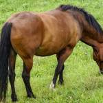
Historical Review of Gastric Ulcers
In the past 30 years, the understanding of equine gastric ulcer disease (EGUD) has advanced by leaps and bounds. Decades ago, though, researchers were just piecing together the “hows” and “whys” of the disease and attempting to arrive at a cure. One prominent researcher involved in crafting a cure for EGUD was veterinarian Al Merritt, who shared his experiences in an interview with Kentucky Equine Research. Extracts of that conversation follow. The complete transcript of the interview can be found in the proceedings of the 2018 Kentucky Equine Research Conference.
When did you first begin to study or research gastric ulcers? At that time, what were the primary research interests in the field? What was happening?
My initial attraction to equine gastric ulcers was in the early 1980s, when I was at the University of Florida, but it was gastroduodenal ulcer disease (GDUD) in foals, an entirely different problem from that seen in adults, which stoked the interest.
I think that the seminal report that brought the attention of gastric ulcers in mature horses was the research concerning necropsy findings of gastric lesions confined to the squamous mucosa, but with no accompanying duodenal lesions, in retired Thoroughbreds in Hong Kong
In those early years, what was considered state-of-the-art technology when it came to diagnosing ulcers? Was diagnosis based solely on clinical signs? What were those signs?
Once we knew gastric lesions could occur in horses, the push was on to apply endoscopy to make a definitive diagnosis. At that time, state-of-the-art technology in human medicine was fiber-optic endoscopy but, except for some colonoscopes which were very expensive, none of the commercial units was long enough for use in the horse. Nonetheless, various manufacturers soon had either units designed for humans, or prototypes being designed for horses, that were at least two meters in length that started to become available, but they still were expensive.
As technology moved from fiber-optic to digital imaging, longer scopes became commercially available, though still expensive, and improved markedly the imaging that could be easily stored in an accompanying processor or a computer for later analysis. Still, the focus early on was on lesions within the squamous mucosa for which a two-meter-long unit sufficed. This is the classic “gastric ulcer disease” described in the horse at that time, seen with high prevalence in animals in training. As more and more animals were scoped, however, it became clear that lesions could also occur within the glandular mucosa and that required the need of longer scopes for detailed observation. Most videoendoscopes now used in horses are three meters long, which allows for close inspection of the upper duodenum as well as the full stomach.
With respect to clinical signs, recognition of those that could be indicative of gastric ulcer disease resulted more from scoping animals that just “weren’t right” than the other way around. That is, complaints of horses showing uncharacteristic irritability, failure to finish a meal, signs of mild abdominal discomfort, and failure to perform up to expectation were grounds for gastric endoscopy, and many of those animals had significant ulceration, most commonly of the squamous region. But as more and more endoscopies have been done over time for one reason or another, we now know that quite severe squamous lesions can also be present in animals that appear clinically normal.
Can you clarify the terminology for horse owners?
As indicated above, of major clinical interest in gastric ulcers in adults during the late 1980s and most of the 1990s was those lesions occurring in the squamous mucosa and their effect on performance, especially since they seemed to have greatest prevalence in animals in training. Lesions of the glandular mucosa that were also seen from time to time were attributed little clinical significance except for those few that were associated with intensive NSAID therapy. Then, in 1999, a group of academic and private clinicians, of which I was one, who had been assembling annually for a number of years to share ideas and current research results concerning gastric ulcers under the auspices of Merial, published a consensus report regarding the current knowledge of the problem.
That report suggested classifying all of the various conditions that manifest as ulcerative lesions within the esophagus, stomach, or upper duodenum—sites with potential exposure to gastric acid—as “equine gastric ulcer syndrome” (EGUS). Further explanation of the term stated, “While the name does not adequately describe all manifestations of the syndrome, adaptation into conventional vocabulary suggests that the reference be maintained.” From this, the EGUS term took off. However, it was often used with no qualification of whether it was referring to squamous or glandular lesions, or both, which implied that EGUS is one disease rather than it being a term that refers to a number of unique problems that manifest upper gastrointestinal ulceration.
Thus, in 2009, I wrote an editorial in the Equine Veterinary Journal titled “Appeal for the Proper Use of the Term “EGUS”: Equine Gastric Ulcer Syndrome,” in which I stated that it is important to include a qualifier concerning which particular form of problem falling within the EGUS domain is under consideration in any written or oral communication. “Only this will reinforce the very important concept that EGUS is not one disease, which is critical to promoting further accurate knowledge about these problems in the upper GI tract of the horse.” That appeal was gratefully taken to heart by many of the current workers in the field, most notably Ben Sykes who was instrumental, along with some European colleagues, in publishing in 2015 in Journal of Veterinary Internal Medicine a consensus statement from the European College of Equine Internal Medicine promoting two subclassifications of the EGUS term: equine squamous gastric disease (ESGD) and equine glandular gastric disease (EGGD).
In those days, what were the standard treatments for gastric ulcers?
Once EGUS was recognized, it was only natural to turn to therapeutic protocols similar to those being used in human medicine, most notably agents that either buffered or blocked secretion of gastric acid. It was pretty clear early on that effective antacid therapy promoted healing of ESGD and this was somewhat good fortune given what we now know about the major role of gastric acid in the pathogenesis of squamous ulceration, and the similarities between ESGD and human gastroesophageal reflux disease (GERD), for which the prime treatment is an antacid. Histamine-2 receptor antagonists were the antisecretory drug class of choice in human medicine in the 1980s and those that were available were, of course, formulated for human use and dosage. Were they effective in the horse, and at what dose?
In 1984, a veterinarian developed a technique for chronic gastric cannulation of the horse that allowed collection of gastric contents from conscious animals over various time periods. With that model, we reported that intravenous ranitidine effectively blocked acid secretion for a number of hours. But the intravenous route was certainly not practical for routine use, so homemade preparations of ground-up pills or powder in some vehicle like commercial syrup were devised, and through various studies and evidence-based experience, a dose of 6.6 mg/kg of ranitidine three times a day was settled upon, considerably higher than that recommended for humans
Other agents tried early on included the PGE2-inducing compound sucralfate and other substances that presumably provided a protective coating of the squamous mucosa against acid damage, but these never took hold in a big way. The same for oral-buffering agents, which we later showed, in cannulated animals, had only a very short-term effect.
Then came omeprazole, with the first studies of acid inhibitory effect in cannulated horses reported in 1992. We subsequently worked with Merial to develop an oral paste formulation of omeprazole that was finally marketed commercially. While this product was considerably more expensive than ranitidine, who could argue against a paste formulation that only needed to be given once a day? The rest is history.
Further information from this presentation and others can be found in the Proceedings of the 2018 Kentucky Equine Research Conference.
Most Popular
 Putting Weight on a Skinny Horse (326,955)
- Benefits of Beet Pulp for Horses (217,433)
 Hot Blood, Warm Blood, Cold Blood in Horses (174,499)
 Possible Link Between Selenium and Cribbing in Horses (163,853)