Inflammatory Bowel Disease in Horses

Diagnosing and treating inflammatory bowel disease (IBD) in horses can be challenging for veterinarians and horse owners.
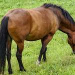
As in other species, including humans, IBD occurs due to the influx of inflammatory cells into the walls of the small and large intestine. The physical presence of these cells causes inflammation and malabsorption of dietary nutrients, which explains why many horses suffer weight loss despite having a normal appetite. A combination of factors is thought to contribute to the development of IBD, such as genetics, status of the intestinal microbiome, and environment.
Several forms of IBD exist, and the best diagnostic technique reported to date involves obtaining biopsies of the small intestinal (duodenum) or large intestinal wall through an endoscope. Analysis of these biopsies can reveal granulomatous enteritis, multisystemic eosinophilic epitheliotropic disease, diffuse eosinophilic enterocolitis, proliferative enteritis, and lymphocytic-plasmacytic enterocolitis.
One veterinary researcher noted,* “Enteral biopsies may be a useful diagnostic aid in the workup of horses thought to have IBD, however further research is required to demonstrate their true diagnostic value. There is need for standardization of the biopsy procedure and of the histopathological interpretation protocol. It is important to combine biopsy results with all other applied diagnostic tests during the workup of a horse thought to have IBD and not to overestimate the diagnostic and prognostic value of any diagnostic test.”
Another potential method of diagnosing IBD involves conducting an oral glucose tolerance test, a procedure similar to the one used to help diagnose insulin dysregulation in horses. The theory is that IBD causes malabsorption; therefore, less sugar than expected would be absorbed systemically in horses with IBD compared to healthy horses.
Most horses with IBD present with:
- Recurrent colic;
- Weight loss, often despite a healthy appetite;
- Poor performance and lethargy; and
- Ventral edema (fluid accumulation in the dependent parts of their bodies such as the underside of the abdomen and lower limbs).
Even without a failsafe test for diagnosing IBD, performing some diagnostics remains important, at least to rule out other causes of chronic diarrhea, including:
- Chronic parasitism (cyathostomosis, large strongyle infection, other intestinal parasites);
- Salmonellosis;
- Equine proliferative enteropathy (Lawsonia intracellularis);
- Ulcerative colitis (Rhodococcus equi);
- Peritonitis;
- Idiopathic colonic dysfunction;
- Nonsteroidal anti-inflammatory drug toxicity (right dorsal colitis);
- Sand enteropathy;
- Equine gastric ulcer syndrome, and a large variety of less common causes.
In many cases of diarrhea, including inflammatory bowel diseases such as lymphocytic-plasmacytic enterocolitis, the prognosis is reportedly guarded at best.**
Although not demonstrated in controlled clinical studies in horses, omega-3 fatty acids have yielded some benefit in a number of human studies for patients suffering various forms of inflammatory bowel disease.*** Kentucky Equine Research offers EO-3, a potent marine-derived oil that is rich in both EPA and DHA. This product is top-dressed onto feed and supplies almost 10,000 mg of omega-3 fatty acids per 30 mL serving.
*Boshuizen, B., M. Ploeg, J. Dewulf, et al. 2018. Inflammatory bowel disease (IBD) in horses: A retrospective study exploring the value of different diagnostic approaches. BMC Veterinary Research. 14(1):21.
**Oliver-Espinosa, O. 2018. Diagnostics and treatments in chronic diarrhea and weight loss in horses. Veterinary Clinics of North America Equine Practice. 34(1):69-80.
***Limketkai, B.N., A. Wolf, A.M. Parian. 2018. Nutritional interventions in the patient with inflammatory bowel disease. Gastroenterology Clinics of North America. 47(1):155-177.
Most Popular
 Putting Weight on a Skinny Horse (327,084)
 Benefits of Beet Pulp for Horses (217,577)
 Hot Blood, Warm Blood, Cold Blood in Horses (174,559)
 Possible Link Between Selenium and Cribbing in Horses (163,889)