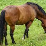
Muscle Atrophy in Horses
When a muscle of a horse decreases in size, seemingly melting away, it’s known as muscle atrophy. When this occurs, the primary concerns to a horse owner are whether the muscle will regenerate, returning to its normal size and shape, and how to treat the horse to help it recover.
According to Stephanie J. Valberg, D.V.M., Ph.D., of the University of Minnesota College of Veterinary Medicine during her presentation at the 2011 Texas Equine Veterinary Association conference, obtaining a thorough history and performing a complete physical, neurologic, and lameness examination are critical once muscle atrophy has been identified.
The history should include any injuries, falls, vaccinations, medications, environmental problems, signs of disease, and foaling records.
Valberg said, “From the historical information, it should be apparent whether there is focal, multifocal, or generalized atrophy; whether the horse is losing muscle mass in spite of a good appetite; and whether there is any association between atrophy and lameness, neck or back pain, trauma, or concurrent disease.”
The physical examination includes palpation of muscle masses, observation of muscle mass symmetry, and notation of heat, pain, swelling, or other abnormalities in a muscle. Valberg said a thorough lameness examination should be performed, and explained that any sign of neurologic disease should be followed up by a complete neurologic exam.
“In terms of prognosis and treatment options, it is often helpful to determine if atrophy is due to wasting of the muscle cells (myogenic atrophy) or wasting due to damage to the nerves supplying the muscle (neurogenic atrophy),” noted Valberg.
She said veterinarians often use one or more of the following tests to assist in prognosis and treatment options:
Diet inspection. The amount and type of feed the horse is actually eating as well as quality of green pasture, which contains vitamin E, should be determined.
Muscle enzymes. Myogenic atrophy is often associated with elevations in serum creatine kinase (CK) activity and if rhabdomyolysis is more than 6-12 hours in duration, elevations in aspartate transaminase (AST) and lactate dehydrogenase (LDH) may also be present.
Vitamin E concentration. Vitamin E concentration can be measured in serum samples; however, variability in serum levels can be quite large and pooling of several samples is often recommended to accurately assess deficiency.
Ultrasonography. Diagnostic ultrasonography can be useful in identifying the extent and depth of focal muscle atrophy as well as cervical facet lesions and pelvic fractures, noted Valberg. She emphasized that the appearance of muscle on ultrasound can change due to the way the horse is standing and whether the muscle is under tension, so it is important that the horse is standing squarely and bearing weight evenly.
Radiography. Horses with focal areas of cervical (neck) muscle atrophy should have radiographs obtained of the cervical spine to look for areas of osteoarthritis that might impinge upon the cervical motor nerves.
Nuclear scintigraphy. Valberg said this modality is useful for identifying pelvic and spinal fractures that cannot be seen on radiographs. Those fractures can cause damage to motor nerves near the injury and result in neurogenic atrophy.
Electromyography (EMG). Although not readily available in most veterinary practices, Valberg said EMG testing allows the extent of muscle atrophy to be examined and provides information that might be diagnostic for the cause of muscle atrophy. “Horses with abnormalities in the electrical conduction system of muscle or denervation of motor units show abnormal spontaneous electrical activity,” reported Valberg.
Muscle biopsy. “Evaluation of muscle fiber sizes, shapes, and types in a biopsy sample can determine if regeneration can be expected or if there is neurogenic atrophy that might respond well to therapeutic intervention such as vitamin E, equine protozoal myeloencephalitis (EPM) treatment, or rehabilitation therapy,” explained Valberg, whose lab at the University of Minnesota offers several types of muscle biopsy services. Submission information can be obtained here.
She said other tests to consider would include dexamethasone suppression test for Cushing’s disease, compete blood count (CBC), complete chemistry profile, cerebrospinal tap, and intestinal absorption studies for systemic wasting diseases.
Muscle Wasting Problems and Prognoses
Myogenic atrophy. Valberg mentioned that 1-5% of muscle mass undergoes remodeling on a daily basis. “If a negative nitrogen balance occurs, net protein withdrawal from the skeletal muscle mass begins within 48 to 72 hours,” she said. “With malnutrition, 30-50% of the muscle mass may be lost in the first 1 to 2 months. Some myogenic atrophy problems are caused by malnutrition, disuse atrophy, Cushing’s disease, secondary to severe rhabdomyolysis, polysaccharide storage myopathy (PSSM), and immune-mediated myopathy.
Neurogenic atrophy. “Denervation removes the normal low-level tonic neural stimulus that is necessary to maintain muscle fiber mass,” stated Valberg. “Complete denervation of a muscle results in more than a 50% loss of muscle mass within a two- to three-week period.” Some causes of neurogenic atrophy include equine motor neuron disease (EMND), EPM, cervical vertebral osteoarthritis, thoracic, lumbar, or pelvic fracture, or post-anesthetic myoneuropathy.
Treatment Options
Valberg discussed treatment options that are available for some of the problems that cause muscle atrophy: Cushing’s disease (pergolide), PSSM (diet and exercise modification), EPM (ponazuril), EMND (vitamin E), cervical vertebral osteoarthritis (corticosteroid injection of articular facets).
Valberg noted that if the muscle biopsy does not show extensive fibrosis in cases of disuse or neurogenic atrophy, physical therapy might be effective in regenerating muscle. Some techniques she mentioned included deep-heating ultrasound, electroacupuncture, electrical muscle stimulation, resistance training in water (AquaPacer treadmill), and work on hills or sloped treadmills.
“We have had excellent results in regenerating atrophied muscles using physical therapy,” she added.
Most Popular
 Putting Weight on a Skinny Horse (327,041)
 Benefits of Beet Pulp for Horses (217,528)
 Hot Blood, Warm Blood, Cold Blood in Horses (174,537)
 Possible Link Between Selenium and Cribbing in Horses (163,873)