Preventing and Treating Bone Cysts in Horses
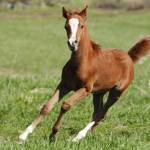
In addition to causing lameness in young horses, disturbing training schedules, and leading to osteoarthritis later in life, bone cysts are challenging to treat.
“Subchondral bone cysts are fluid-filled and often occur at or near weight-bearing surfaces, interrupting the normal pain-free and frictionless transfer of weight between long bones,” explained Laura Petroski-Rose, B.V.M.S, a Kentucky Equine Research veterinarian.
Subchondral bone cysts usually form during the development of the foal’s skeletal system. Because of this, they are considered a developmental orthopedic disorder. The two prevailing theories for cyst development include: (1) failure of proper ossification of bone in foals; and (2) subchondral bone trauma, often during exercise.
Both processes result in inflammation, contributing to clinical signs of disease (heat, pain, swelling), and possible enlargement of the cyst with the passing of time.
Treatment options vary from conservative methods such as stall rest, anti-inflammatory drugs, and intra-articular medications to more invasive surgical techniques.
“In surgical cases, the goal is to remove the entire contents of the subchondral bone cyst, including the lining. Any remaining fragment could result in continued inflammation, lameness, and poor performance,” Petroski-Rose warned.
As recently described*, however, surgery requires the veterinarian to accurately identify where exactly the three-dimensional cyst exists inside the bone to ensure complete excision. Because radiographs—one of the most common methods used to diagnose subchondral cysts—are two-dimensional images of three-dimensional structures, this is sometimes difficult.
To help surgeons better identify the exact location of subchondral bone cysts, Jackson and colleagues from Switzerland recently proposed using an “aiming device” and computed tomography. Together, these devices reportedly allow precise debridement of subchondral bone cysts, thereby decreasing chances of residual cyst tissue and optimizing surgical outcomes.
“Ideally, developmental orthopedic disorders would be minimized by ensuring optimal nutrition of the foal. However, because trauma, genetics, and environmental factors also contribute to this musculoskeletal condition, subchondral bone cysts can’t always be avoided. If your horse does undergo surgery, consider using a joint supplement to minimize the chances of osteoarthritis later in life,” advised Petroski-Rose.
Kentucky Equine Research offers several high-quality joint supplements, including Synovate HA with hyaluronic acid, the glucosamine- and chondroitin sulfate-containing product KER-Flex, and the marine-derived fish oil supplement, EO-3. Following surgery, support bone health and strength with Triacton. Look for Glucos-A-Flex in Australia, too.
*Jackson, M.A., S. Ohlerth, and A.E. Fürst. 2019. Use of an aiming device and computed tomography for assisted debridement of subchondral cystic lesions in the limbs of horses. Veterinary Surgery 48(S1):15-24.


