Use of Ultrasound in Diagnosing Osteochondral Defects in Horses
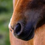
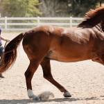
Cartilage defects in joints may shorten the performance careers of horses. Diagnosing cartilage lesions early in a horse’s career can hasten treatment, allowing affected horses to remain just as competitive as those without cartilage defects. While ultrasonography has the potential to serve as a tool to help diagnose cartilage defects, available data do not support its use in a clinical setting currently.
“Prior to being sold, many young horses, especially those destined for high-performance careers, undergo routine radiography of joints. Veterinarians look for cartilage lesions indicative of osteochondrosis in high-use joints, including stifles, hocks, and ankles. Depending on their severity and location, these lesions may preclude the sale of the horse and raise welfare concerns for its future,” explained Kathleen Crandell, Ph.D., a nutritionist for Kentucky Equine Research.
The underlying cause of osteochondral lesions remains unclear, but one theory involves failure of endochondral ossification, the process that forms bone from cartilage. As a result of this failure, the layer of bone underlying the cartilage develops cystic lesions filled with synovial fluid. In some instances, the overlying cartilage breaks away from the lesion and floats freely within the joint.
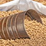
“Failure of endochondral ossification is classified as a developmental orthopedic disease, occurring in growing horses. While multifactorial in nature, the nutritional factors believed to contribute to osteochondrosis include rapid growth rate, high-carbohydrate diet, and mineral imbalances,” Crandell said.
“Strategies for preventing osteochondral lesions include controlling the amount and type of feed as well as checking for the appropriate balance of nutrients,” she continued. “Using software such as Gro-Trac to monitor growth rate can help identify growth abnormalities as they are happening. This, in turn, may lead to management strategies aimed at manipulating growth safely.”
To support healthy bone growth and density, Crandell recommends supplementing horses with Triacton. This nutritional supplement contains highly bioavailable minerals and vitamins essential for sound skeletal development and maintenance, including a proprietary source of calcium.
In human medicine, physicians once relied on ultrasound to detect osteochondrosis-type lesions; however, MRI has largely replaced ultrasonography. In horses, veterinarians have only trifled with ultrasonography for diagnosing these lesions. Instead, they remain dependent predominantly on radiographs for diagnosis.
Recognizing the importance of achieving an accurate and early diagnosis of osteochondral lesions in horses destined for competition, an Irish group of veterinary researchers conducted a systematic literature review.* The researchers gathered all of the available studies that evaluated ultrasonographic diagnosis of osteochondral lesions published between 1970 and mid-2021. Studies were included in the systematic review if ultrasound examinations detected osteochondral-like lesions within a joint and if the ultrasound findings were compared to radiography, arthroscopy, computed tomography, MRI, or postmortem examinations.
Only six studies published between 2009 and 2018 met the inclusion criteria and were comprehensively reviewed.
“Surprisingly, despite the widespread availability and application of ultrasonography in the horse industry, little research regarding this imaging modality in diagnosing osteochondral lesions has been performed,” relayed Crandell.
Data extracted from those six studies revealed the following:
- Ultrasound can evaluate both the superficial and deeper cartilage layers as well as the subchondral bone underlying the cartilage. In contrast, arthroscopy can only assess the superficial cartilage surface and may therefore miss deeper lesions;
- The experience level of the ultrasonographer can limit the value of this technique by failing to obtain quality images or to accurately interpret images; and
- Poor agreement was routinely observed between diagnosing osteochondral lesions via ultrasonography and other imaging modalities, including postmortem examinations.
As a result, the research team concluded that there is “no strong evidence confirming the diagnostic accuracy and validity of ultrasonography in the detection of osteochondral lesions.”
*Hoey, S., D. Stokes, H. McAllister, A. Puggioni, and C. Skelly. 2022. A systematic review evaluating the use of ultrasound in the identification of osteochondrosis in horses. Veterinary Journal 282:105825.
Most Popular
 Putting Weight on a Skinny Horse (326,617)
 Benefits of Beet Pulp for Horses (217,165)
 Hot Blood, Warm Blood, Cold Blood in Horses (174,358)
 Possible Link Between Selenium and Cribbing in Horses (163,758)