Equine Neuromuscular Diagnostic Laboratory
Welcome to the Valberg Neuromuscular Diagnostic Laboratory (NMDL). The NMDL is dedicated to providing the most accurate diagnosis and optimal treatment of muscle disorders in horses.
Percutaneous Needle Biopsy (Middle Gluteal Muscle)
Please download the Percutaneous Needle Biopsy Technique PDF for reference.
Semimembranosus/Semitendinosus
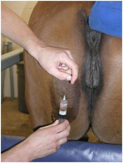
The best site for a semimembranosus biopsy is midway between the tuber ischii and the origin of the Achilles tendon at about the level of the vulvar lips. Avoid tendinous insertions. This site hides scarring under tail hairs and is easily treated if dehiscence occurs.
Sacrocaudalis
The best site for a sacrocaudalis biopsy is 1/2 inch above the origin of the tail and approximately 1/4 inch off midline. Skin retractors are highly recommended for this site.
Procedure
- Lidocaine is injected under the skin but not into the muscle belly. This area has many nerves so 5-7 ml of lidocaine may be necessary.
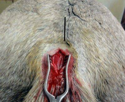
- A 2-inch incision is made through the skin and subcutaneous fat and fascia. The skin is not flexible in this area so a larger skin incision is needed.
- Use skin retractors for visualization.
- Parallel longitudinal incisions are made in the muscle 1/4 inch apart.
- The cranial aspect of the muscle is grasped with forceps and the muscle is dissected out 1/4 inch deep and 1/2 inch long. Don’t pull this muscle out via forceps as that squishing creates atrophy.
- This area can have a lot of subcutaneous fat. Make sure the sample is deep enough to obtain muscle tissue.
Percutaneous Needle Biopsy (Middle Gluteal Muscle)
Please download the Percutaneous Needle Biopsy Technique PDF for reference.
Surgical Biopsy
Semimembranosus/Semitendinosus
The best site for a semimembranosus biopsy is midway between the tuber ischii and the origin of the Achilles tendon at about the level of the vulvar lips. Avoid tendinous insertions. This site hides scarring under tail hairs and is easily treated if dehiscence occurs.
Sacrocaudalis
The best site for a sacrocaudalis biopsy is 1/2 inch above the origin of the tail and approximately 1/4 inch off midline. Skin retractors are highly recommended for this site.
Surgical Procedure
- Lidocaine is injected under the skin but not into the muscle belly. This area has many nerves so 5-7 ml of lidocaine may be necessary.
- A 2-inch incision is made through the skin and subcutaneous fat and fascia. The skin is not flexible in this area so a larger skin incision is needed.
- Use skin retractors for visualization.
- Parallel longitudinal incisions are made in the muscle 1/4 inch apart.
- The cranial aspect of the muscle is grasped with forceps and the muscle is dissected out 1/4 inch deep and 1/2 inch long. Don’t pull this muscle out via forceps as that squishing creates atrophy.
- This area can have a lot of subcutaneous fat. Make sure the sample is deep enough to obtain muscle tissue.
Stephanie Valberg, D.V.M., Ph.D., Dipl. ACVIM, ACVSMR
Dr. Stephanie Valberg is an international leader in diagnosing and treating equine neuromuscular disorders. The overarching goal of Valberg’s research and clinical work is to define the basis for neuromuscular disorders in horses, develop accurate, minimally invasive diagnostic tests, and produce optimal methods for preventing or managing performance limiting diseases.
Note: We do not perform genetic testing. Please submit to the Veterinary Genetics Lab at UC Davis.
Information on this site is intended for information purposes for veterinarians to guide in the diagnostic workup of muscle diseases. Owner-diagnosis and owner-treatment of horses is not recommended and may indeed be dangerous. Owners must consult their veterinarian before implementing any of the treatment recommendations described.
About the Valberg Neuromuscular Diagnostic Laboratory
The NMDL is dedicated to providing the most accurate diagnosis and optimal treatment of muscle disorders in horses.