Middle Ear Disease in Horses
Ask any horsemen to point out the throatlatch, and he will indicate the junction of the head and neck. Ask any horsemen to describe the complicated arrangement of bony and soft tissues assembled in close proximity, and a quizzical look may come over him. Dysfunction of any body complex can occur, and this area, including the temporohyoid joint, is no exception.
The hyoid apparatus is a collection of small bones that supports the larynx, pharynx, and tongue. The hyoid apparatus and the temporal bone connect to form the temporohyoid joint, which lies near the more familiar temporomandibular joint (TMJ). This is a “busy” location as many tissues, blood vessels, and nerve channels intersect here, and disease of the temporohyoid joint can therefore present in many ways.
“Disorders of the temporohyoid joint are collectively referred to as middle ear disease,” explained Catherine Whitehouse, M.S., a Kentucky Equine Research advisor.
Dysfunction of the temporohyoid joint is referred to as temporohyoid osteoarthropathy (THO) and encompasses diseases of the middle ear, the temporohyoid joint proper, the stylohyoid bone, and the base of the skull.
The exact cause of THO is not well understood, though inflammation and infection of the inner or middle ear and guttural pouches have been implicated, as have infection and trauma.
Clinical signs associated with THO include:
- Eyelid paralysis (ptosis);
- Drooping of the muzzle;
- Head tilt;
- Ataxia (incoordination or imbalance);
- Auditory losses; and
- Difficulty swallowing.
In performance horses, THO may manifest as a reluctance to take the bit or carry the head in the desired position.
“A group of Japanese researchers* recently suggested that THO may also be associated with cribbing in horses,” shared Whitehouse.
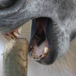
Cribbing is a stereotypical behavior that involves the horse grasping a flat surface with its incisors and producing a grunting noise. This repetitive behavior could subject the hyoid apparatus to excessive pressure, potentially contributing to THO. Cribbing, like other stereotypies, causes damage to facilities and may be linked to certain types of colic.
Diagnosis of THO is typically confirmed through skull radiography, upper airway endoscopy, or computer tomography (CT) scans. Extremely detailed information can be derived from CT scans, revealing changes to the hyoid apparatus, skull, and inner ear. Information from these scans can guide surgical decisions.
The surgical option of choice for THO is a ceratohyoidectomy, which involves removal of small bones within the hyoid apparatus. The procedure reduces the force applied from the hyoid apparatus to the skull and decreases the risk of fracture, and assuages discomfort caused by a diseased joint.
Most horses recover from surgery with few complications, though some horses may have slight nerve deficits that persist after surgery, such as a head tilt or lip droop. Prognosis for full recovery depends largely on the severity of the disease at the time of surgery.
“Nutritional supplements containing glucosamine, chondroitin sulfate, and other ingredients designed to support joint tissues can be offered to horses prior to any trauma or wear-and-tear that could contribute to degeneration of the temporohyoid joint. Examples of such products include KER-Flex, Synovate HA, and the omega-3 product EO-3, previously demonstrated to help horses with osteoarthritis,” relayed Whitehouse.
*Saito, Y., and T. Amaya. 2019. Symptoms and management of temporohyoid osteoarthropathy and its association with crib-biting behavior in 11 Japanese Thoroughbreds. Journal of Equine Science 30:81-85.

