An Overview of Sesamoiditis in Horses
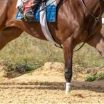
If you’ve been around high-performance horses long enough, you’ve likely heard of sesamoiditis, a cause of unsoundness that can knock out the most hard-knocking athletes.
At the back of each fetlock joint lies two triangular structures called proximal sesamoids. Strictly speaking, each forelimb contains a third sesamoid, called the distal sesamoid or navicular bone, and each hind limb contains a fourth, the patella.
These structures are considered intercalated bones because they arise from within tendons and ligaments and allow for smooth movement and dissipation of focal pressure between the tendon or ligament and the joint that lies beneath.1
By definition, sesamoiditis is inflammation of a sesamoid and its ligamentous attachments, specifically “inflammation of the suspensory ligament at the point where it anchors into the bone.”2 While sesamoiditis occurs in both fore and hind legs, it is more prevalent in forelegs.
The cause of sesamoiditis remains incompletely understood. One study discounted vascular compromise as a trigger, though bone strain in areas of ligamentous attachment may lead to deeper bone inflammation.
Sesamoiditis occurs in all types of athletic horses because the suspensory apparatus bears a significant burden during exercise, yet injuries seem to be most abundant in those that work at speed, such as Thoroughbred and Standardbred racehorses.
If the suspensory ligament is injured extensively at the point of insertion to the proximal sesamoids, the structure is forever changed during healing, as scar tissue forms and the apparatus will function suboptimally when compared to uninjured tissues, often resulting in decreased performance.
Diagnosis of sesamoiditis is made through clinical signs and radiography. Horses may show pain in response to palpation or distal limb flexion test, though lameness can usually be observed after intense exercise. Sesamoiditis can be verified with radiographs, typically oblique views of the fetlock joint.
Treatment typically involves rest and measures to relieve inflammation and pain. Management strategies such as post-workout icing and a reworking of the training schedule to include other exercise, such as swimming, seems to help some horses.
Aside from older horses in active training, sesamoiditis is a common radiographic finding of front and hind fetlocks in Thoroughbred sales yearlings. In one study of 487 yearlings, 127 horses (26%) showed signs of sesamoiditis in survey radiographs.3 In a more recent study of survey radiographs from 318 Thoroughbred yearlings, 24% had evidence of sesamoiditis, and by the time the sales occurred months later, 16% had evidence of sesamoiditis.4
1Durham, M., and S.J. Dyson. 2003. Applied anatomy of the musculoskeletal system. In: M.W. Ross and S.J. Dyson, editors, Diagnosis and Management of Lameness in the Horse. Saunders, St. Louis, MO. p. 86.
2Bramlage, L.R. Operative orthopedics of the fetlock joint of the horse: Traumatic and developmental diseases of the equine fetlock joint. In: Proc. Annual Convention of the American Association of Equine Practitioners 55:96-143.
3Spike, D.L., L.R. Bramlage, B.A. Howard, R.M. Embertson, and S.R. Hance. 1997. Radiographic proximal sesamoiditis in Thoroughbred sales yearlings. In: Proc. Annual Convention of the American Association of Equine Practitioners 43:132-133.
4Pagan, J.D., M. Garzillo, S. Caddel, and E. Phethean. 2020. Relationship between foal growth survey radiographic findings and sales and racing performance in Thoroughbred racehorses: A pilot study.

