Early Detection of Osteoarthritis in Horses
When it comes to screening for osteoarthritis, there’s a new game in town: infrared (IR) spectroscopy. Based on preliminary data, IR spectroscopy can accurately differentiate horses with experimentally induced osteoarthritis from controls, making this an interesting technique worthy of further study.*
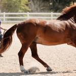
Osteoarthritis is a common degenerative condition of joints that negatively affects performance and quality of life.
“Being able to diagnose OA in the early stages allows us the opportunity to try and slow disease progression and prolong athleticism and patient comfort,” explained Kathleen Crandell, Ph.D., a nutritionist for Kentucky Equine Research.
She adds, “Current means of supporting horses’ joints include injectable polysulfated glycosaminoglycan (PSGAG), injectable hyaluronan (HA), and oral supplements containing chondroitin sulfate, glucosamine, hyaluronic acid, and methylsulfonylmethane. Recent work has also shown the benefits of DHA and EPA in reducing joint inflammation.”
Scientists studying osteoarthritis are looking at the disease through various lenses, each hoping to find reliable and accurate ways of identifying joints in the early stages of the disease. One such approach is analyzing synovial fluid using “omics” technology, such as proteomics. This involves analyzing the proteins in the synovial fluid to determine if certain proteins are overproduced or underproduced in joints with osteoarthritis compared to healthy joints. The “omics” tests, however, are costly and have so far produced inconsistent results, potentially because biomarkers may be intermittently expressed or not proportional to the burden of disease.
A group of veterinary researchers from Colorado State University, New Zealand, Hong Kong, and Canada recently collaborated on a project that assessed the ability of IR spectroscopy to differentiate between joints with experimentally induced osteoarthritis and healthy joints. IR spectroscopy is a laboratory technique that can analyze the molecular composition of small volumes of dried synovial fluid, and those molecular structures can be compared between samples.
“This technique does not require the separation of single molecular species associated with disease but instead provides a complex IR signal produced by an array of molecules,” explained the researchers, adding that IR can “evaluate a range of known and unknown biomarkers simultaneously.”
IR spectroscopy has previously been studied in horses with osteochondrosis and naturally occurring traumatic arthritis, as well as in dogs and humans. Those preliminary studies show IR spectroscopy has high accuracy, sensitivity, and specificity (measures of how reliable a test is to diagnose the disease), making it a potentially useful diagnostic and screening tool for osteoarthritis. It is cost-effective, and collecting synovial fluid is minimally invasive.
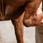
The research team used the well-established “chip fracture” method of experimentally inducing osteoarthritis, a surgical technique that creates a cartilage and bone fragment emulating a traumatic joint injury. A chip fracture was made in one knee (carpal) joint in nine horses by endoscopic surgery, leaving the contralateral knee joint intact (control). In addition, eight horses underwent a “sham” operation in which an endoscope was introduced into one knee joint, but no chip was created. The contralateral knee of the sham-operated horses also served as a control (sham control).
Synovial fluid samples were collected immediately before surgery and weekly for nine weeks. Those samples were then analyzed via IR spectroscopy.
The accuracy for distinguishing between joints with osteoarthritis and the sham-operated, osteoarthritis-control, and sham-control joints was 80%.
“IR spectroscopy accurately discriminates between synovial fluid in joints with induced osteoarthritis and controls,” relayed the researchers.
“While this approach can differentiate affected joints from controls with good accuracy in a research setting, it was a short study, lasting only nine weeks, using an experimental model. Studies using horses with more advanced osteoarthritis and with naturally occurring disease would be beneficial. Further, advances in the IR technique are underway using ultra-broadband quantum IR spectroscopy, which holds promise for increasing sensitivity of the test,” noted Crandell.
Detecting joint changes early in the course of osteoarthritis would allow us to intervene earlier with treatment strategies to slow disease progression. What would be equally beneficial is identifying disease-modifying agents of osteoarthritis that can stop the disease in its tracks or help the joint tissues heal.
Crandell concluded, “Don’t wait until your horse has signs of osteoarthritis to start supplementing with chondroitin sulfate, glucosamine, or omega-3 fatty acids. Research shows that prophylactic administration of these joint supplements may delay the onset of osteoarthritis. This means that owners should offer joint supplements to young, healthy horses prior to joint trauma or natural wear and tear.”
*Panizzi, L., M. Vignes, K.E. Dittmer, M.R. Waterland, C.W. Rogers, H. Sano, C.W. McIlwraith, and C.B. Riley. 2024. Infrared spectroscopy of synovial fluid shows accuracy as an early biomarker in an equine model of traumatic osteoarthritis. Animals (Basel) 14(7):986.
Most Popular
 Putting Weight on a Skinny Horse (331,441)
 Benefits of Beet Pulp for Horses (221,409)
 Hot Blood, Warm Blood, Cold Blood in Horses (176,475)
 Possible Link Between Selenium and Cribbing in Horses (165,368)