Gastrointestinal Tract Basics: The Horse’s Foregut
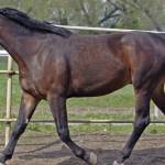
The foregut of the horse includes the esophagus, stomach, and small intestine. The small intestine is further divided into the duodenum, jejunum, and ileum. The foregut of the horse is much like those of other monogastric (simple-stomached) animals such as humans, pigs, and dogs.
The esophagus is a long muscular tube that is collapsed upon itself when it is not carrying feed or water. The esophagus begins in the back of the throat and then travels over the middle of the neck to the left side of the neck where it enters the chest. Once in the chest, the esophagus is found over the heart and windpipe. It then goes through the diaphragm and enters the stomach. The first two-thirds of the esophagus is composed of skeletal muscle (same as the muscle responsible for movement of the body) and the last one-third is made up of smooth muscle (muscle responsible for movement of the gut). The most common problems involving the esophagus are acute obstruction (choke), strictures (narrowing or scarring), and diverticula (outpocketing). The usual sites for choke are the thoracic inlet, where the esophagus enters the chest, and over the base of the heart. The most common diagnostics to evaluate the esophagus are endoscopy and contrast esophagrams, during which the horse is given radiographic contrast material and radiographs are taken to look for strictures or diverticula.
The stomach of the horse is divided into the nonglandular portion that is lined by the same type of cells that make up skin and line the inside of the esophagus, and the glandular portion that produces stomach acid. Unlike other species, the parietal cells in the horse’s stomach continually secrete acid. The margo plicatus is the noticeable division between the glandular and nonglandular stomach. Gastric ulcers most commonly occur in the squamous portion of the stomach along the margo plicatus. Less common conditions that affect the stomach are gastric impaction and cancer (squamous cell carcinoma). Bot fly larvae are commonly seen attached to the squamous portion of the stomach, but they rarely cause any problems, even if present in large numbers. Gastroscopy is the best diagnostic method to examine the stomach.
The first portion of the small intestine after it exits the stomach is the duodenum. It is relatively short, and it is where the pancreatic and bile ducts empty important digestive enzymes. The jejunum is the most extensive portion of the small intestine, and it is the major site of carbohydrate and amino acid digestion. The last section of the small intestine is the ileum, which is important for absorption of certain vitamins and bile acids. Medical conditions that may involve the small intestine include duodenitis/proximal jejunitis, roundworm impactions, tapeworm infections, and ileal impactions. Surgical problems of the small intestine can be very serious and life-threatening. These include small intestinal volvulus (twisted gut), pedunculated lipoma, and small intestinal hernias.


