Needle Arthroscopy for Equine Lameness
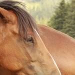
What would you do if radiographs of your horse’s knee revealed no lesion or injury, despite persistent swelling and lameness? Researchers suggest a technique called needle arthroscopy might be the next reasonable option in securing a diagnosis.*
Needle arthroscopy involves inserting a minute endoscope—an illuminated optical instrument—into a joint to thoroughly visualize and evaluate the structure. Unlike much larger, more cumbersome full-sized arthroscopes, needle arthroscopes allow for greater maneuverability within the cramped confines of the joint capsule.
“Needle arthroscopy helps veterinarians diagnose ‘radiographically silent lesions’ of the carpus, or knee, such as certain fractures and tears of intercarpal ligaments,” explained Catherine Whitehouse, M.S., a Kentucky Equine Research advisor.
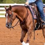
“Whereas magnetic resonance imaging, computed tomography, and traditional arthroscopy all require the horse to be anesthetized and laid down, needle arthroscopy is performed in the standing, sedated horse,” she said, “minimizing complications associated with anesthesia, such as injuries during recovery, and reducing the cost of the procedure.”
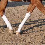
To demonstrate the feasibility and utility of needle arthroscopy, the veterinarians practiced needle arthroscopy in cadaveric limbs before moving on to six sound horses. In each horse, two of the three carpal joints were assessed: the radiocarpal joint (between the forearm and the top of the knee) and the middle carpal joint (between the two rows of carpal bones). While racehorses often sustain injury to the middle carpal joint, pleasure and sport horses more frequently have damage to the radiocarpal joint.
Here’s what the researchers found:
- Each joint took less than 10 minutes to examine;
- Injured structures within the joint were clearly visualized with needle arthroscopy;
- The main complication encountered during needle arthroscopy was excessive movement of the horse despite sedation;
- No complications noted following the procedure;
- The incision that allowed passage of the arthroscope was no longer noticeable 48 hours after surgery; and
- Horses resumed paddock turnout within six days of the procedure.
This study shows that diagnostic needle arthroscopy offers an affordable method of achieving early diagnosis of intra-articular lesions, thus expediting treatment. Addressing equine joint injuries as early as possible will decrease the chances of secondary cartilage damage or joint instability that may threaten the horse’s short- and long-term athletic career.
“With any joint injury, inflammation may initiate the cascade of events that almost inevitably results in the development of osteoarthritis. Oral joint supplements help slow the development of degenerative changes after injury to a joint and may prolong the careers of athletic horses,” advised Whitehouse.
Kentucky Equine Research offers several high-quality supplements designed to protect joints before and after injuries.
“Studies also show that using these products in healthy horses prior to any injury may protect the joint from inflammation and the initiation of degenerative changes,” Whitehouse added.
*Kadic, D.T.N., L. Miagkoff, and A.G. Bonilla. 2020. Needle arthroscopy of the radiocarpal and middle carpal joints in standing sedated horses. Veterinary Surgery 49(5):894-904.
Most Popular
 Putting Weight on a Skinny Horse (328,697)
 Benefits of Beet Pulp for Horses (218,974)
 Hot Blood, Warm Blood, Cold Blood in Horses (175,318)
 Possible Link Between Selenium and Cribbing in Horses (164,434)