Ultrasound Exams in Injured Horses: Take Off the Weight
The musculoskeletal health of performance horses must be considered throughout a horse’s career, even in times of soundness. When something goes wrong with the soft tissues within this system, an ultrasound examination is often recommended. How this ultrasound is performed, however, may make a big difference in achieving a prompt and accurate diagnosis that will drive treatment recommendations.
“For bone and joint injuries, veterinarians most often use X-rays to make a diagnosis. For soft tissues, like tendons and ligaments, ultrasound frequently replaces or follows X-rays,” said Peter Huntington, B.V.Sc., M.A.C.V.Sc., a veterinarian and director of nutrition at Kentucky Equine Research (Australia).
When performing an ultrasound exam of a tendon or ligament, the traditional technique involves having the horse stand, bearing equal weight on the limbs. When an ultrasound does not reveal a clear diagnosis, computed tomography (CT) or magnetic resonance imaging (MRI) may be recommended by your veterinarian.
“Ultrasound examinations are more widely available and cost-effective than CT and MRI, and can be used to monitor response to treatment over time. Therefore, accurately identifying lesions to tendons and ligaments by ultrasound whenever possible would most benefit the horse,” explained Huntington.
Recently, equine researchers* found that lifting the limb being examined can reveal injuries that wouldn’t be observed on a weight-bearing limb.
“According to that study, bearing weight can decrease the size of the lesion in the abnormal soft tissues. It can also decrease the ultrasonographic visibility of certain injuries and obscure fluid within joints or tendon sheaths that could mask an abnormality,” Huntington said.
After examining 62 injured suspensory ligaments, the researchers found that certain abnormalities, such as longitudinal fiber disruption or a “split” along the length of the suspensory ligament, can only be found when the limb is lifted.
“The research team concluded that the new approach of using ultrasound on a lifted limb provides a more accurate representation of the severity of the lesions and allows veterinarians to monitor response to treatment and guide owners in the return to soundness,” summarized Huntington.
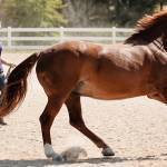
Regardless of which structure in the limb suffers trauma, injured horses often require abrupt stable confinement and lay-up.
“When horses are confined, they rapidly lose bone mineral density, which means they may have an increased risk of bone injury when they resume training. High-quality bone supplements, like Triacton and DuraPlex, increase bone density and prevent bone loss during stable confinement,” said Huntington.
*Werpy, N., K. Chapman, and L. Griffith. 2020. Non-weight bearing ultrasonographic examination allows the diagnosis of longitudinal fiber disruption (split) in equine suspensory ligament branches not visible on weight bearing examination. Veterinary Radiology and Ultrasound 62(1):84-97.
Most Popular
 Putting Weight on a Skinny Horse (328,382)
 Benefits of Beet Pulp for Horses (218,742)
 Hot Blood, Warm Blood, Cold Blood in Horses (175,179)
 Possible Link Between Selenium and Cribbing in Horses (164,361)