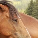
Hands-On Horse Research: Radiographic Photodensitometry In Action
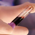
Radiographic photodensitometry measures bone mineral content (BMC) and skeletal strength in horses. This technique is used to monitor the effects of nutrition, exercise, and training.
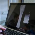
Radiographic photodensitometry is based on the principle that absorption of x-radiation as it passes through bone, and therefore the amount of light passing through a radiograph, is related to the mineral content of the bone. In order to make direct comparisons between x-rays, a reference standard must be exposed simultaneously with the bone.
The reference standard is an 11-step aluminum stepwedge. Aluminum has an atomic number close to hydroxyapatite, the main mineral constituent of bone, and x-rays are absorbed by aluminum to the same degree as bone mineral. When comparisons are made, bone mineral content is expressed as a radiographic bone aluminum equivalent (RBAE), which can be used to calculate actual BMC.
The third metacarpal, or cannon bone, is the preferred site for radiographic photodensitometry. Because it has minimal soft tissue covering, the cannon bone provides a reliable assessment of cortical bone quality.
Radiography also provides an opportunity to make measurements of cortical thickness, medullar cavity width, and bone width. Research has shown that with specific training and nutritional supplements bones can be remodeled in response to exercise, which makes morphometric measurements important.
Learn more about KER’s research program here.
Most Popular
 Putting Weight on a Skinny Horse (328,731)
 Benefits of Beet Pulp for Horses (219,010)
 Hot Blood, Warm Blood, Cold Blood in Horses (175,332)
 Possible Link Between Selenium and Cribbing in Horses (164,441)